 As you raise your glass of champagne tonight and toast this wonderful year of biodiversity, don't forget the parasites. And, to help you remember, today's parasite is after the grapes cultivated for wine. Guignardia bidwellii is a species of ascomycetous fungus that causes a disease called "Black rot" in many varieties of grapes in North America and now Europe, South America, and Asia as well. The vectors of this disease are not mosquitoes nor plant bugs, but rather raindrops, which splash the infective spores onto uninfected plants. Infection of the fruits will cause the grapes to shrivel up into what are known in the industry as "mummies" and these can serve as a good place for the fungus to overwinter.
December 31, 2010
December 31 - Guignardia bidwellii
As you raise your glass of champagne tonight and toast this wonderful year of biodiversity, don't forget the parasites. And, to help you remember, today's parasite is after the grapes cultivated for wine. Guignardia bidwellii is a species of ascomycetous fungus that causes a disease called "Black rot" in many varieties of grapes in North America and now Europe, South America, and Asia as well. The vectors of this disease are not mosquitoes nor plant bugs, but rather raindrops, which splash the infective spores onto uninfected plants. Infection of the fruits will cause the grapes to shrivel up into what are known in the industry as "mummies" and these can serve as a good place for the fungus to overwinter.
December 30, 2010
December 30 - Bunocotyle progenetica
 Bunocotyle progenetica is another parasite (see also Parvatrema margaritense) that has been thoroughly studied in the White Sea. Being a hemiurid trematode, it possesses all the typical life cycle stages. But when it comes to hosts, we see something entirely different. Hydrobia snails serve as an “all-in-one” habitat throughout the parasite's life. That is, cercariae don't leave the rediae but instead continue their development, up to an adult stage, still inside the same mollusc. The photo shows a redia with adults inside, with visible eggs inside them. The eggs are transferred to neighboring Hydrobia molluscs after the host's death. This favours increased snail exploitation by B. progenetica, since it doesn't require the host to live long. Thus the entire life of B. progenetica passes inside its host, with no free-living stage at all. This phenomemon is not uncommon among parasites as it provides maximum protection against a potentially hostile environment. The serious drawback of such a strategy, though, is lack of dispersal opportunities. It's possible to overcome this by using mobile hosts, however, not the case here, thus B. progenetica is a good example of just how odd parasites can sometimes be.
The PhD thesis referenced is entirely dedicated to this parasite, while the second paper only has certain comments on it.
Levakin I.A. Realization of a one-host life cycle of Bunocotyle progenetica (Trematoda: Hemiuroidea: Bunocotylinae) at the White Sea intertidal zone. PhD thesis manuscript, 2007. (In Russian)
Gorbushin, AM, 1997: Field evidence of trematode-induced gigantism in Hydrobia spp. (Gastropoda: Prosobranchia). J. Mar. Biol. Ass. UK 77 , 785–800.
Contributed by Anya Gonchar, photo by Ivan Levakin.
December 29, 2010
December 29 - Eremitilla mexicana
 Back in 1985, Wayt Thomas, a scientist from the New York Botanical Garden discovered an unusual plant in Mexico. It had a little bloom of dense flowers that kind of looked like a pinecone and nothing else but a thick stalk - no leaves or chlorophyll anywhere. It was so unusual that Thomas did not know what it was and could only speculate as to even what family it might be in. The strange plant eventually made its way to George Yatskievych at the Missouri Botanical Garden and twenty years after it was first discovered, he traveled back to Mexico in search of more. He went to the same location - and even employed the very same guide that Thomas had - and finally, after several days of hunting through stream beds in the Sierra Madre del Sur, they found a small population and took a few samples and many photographs. They did not collect very many because it is believed to only occur in this one small region - it has never been observed elsewhere. A second trip allowed Yatskievych to identify the host plants as Hedyosmum mexicanum and it has now been named Eremitilla mexicana, which means "little Mexican hermit."
Photo by George Yatskievych.
December 28, 2010
December 28 - Hyalomma dromedarii
 The three wise men are said to have brought three gifts, but perhaps they brought four. The tick, Hyalomma dromedarii, is the most common ectoparasite of camels found in the Middle East. Because of the high temperatures, the females need to burrow down into the sand to lay their eggs. The larvae find a host and feed, but unlike ticks in more temperate climates that usually then drop off to molt, the larvae of H. dromedarii stay put on their host, molt, and feed again. The first host may be a rabbit, hedgehog, bird, or other small livestock, however if the first host that they feed from is a camel itself, they will sometimes stay right there and complete their entire life cycle on the same host. Dropping off into the hot sand is just far too risky, it seems.
Image is from this site.
December 27, 2010
December 27 - Macrophomina phaseolina
 One of the gifts that the Three Wise Men brought was frankincense, which is derived from the resin of the tree Boswellia serrata. While frankincense has been considered as a remedy for many different types of infectious diseases, B. serrata itself is by no means free from the scourge of infection itself and is plagued by the fungus Macrophomina phaseolina, which causes the disease known as Charcoal Root Rot. This fungus infects more than 300 species of plants, and can cause high mortality among tree seedlings. Macrophomina phaseolina survives and overwinters as small, black spores (call microsclerotia), hidden in the soil or debris from previously infected plants. When a growing root of a plant encounters a dormant spore, it germinates and begins growing all over the root and penetrating into the root cortex. From there, the fungus penetrates through the cortex and inner bark and into the taproot. The infected seedling eventually dies from the gradual destruction of its root system. Just prior to the death of the host, the M. phaseolina produces spores that are deposited in the inner bark of the lower stem and roots. When the host eventually dies and decays, the spores are released into the soil where they wait for an encounter with yet another growing seedling.
Contributed by Tommy Leung.
December 26, 2010
December 26 - Plasmodium vivax
 In Christian lore, three wise men, the magi, traveled from the East bearing gifts for the baby Jesus. These gifts were gold, myrrh and frankincense, a resin made from trees in the genus Boswellia. The reason for the gold seems obvious, myrrh was used as an incense, which had to have made the stable smell better, and frankincense was used for many things, several related to improving ones health, including ingesting the resin to combat arthritis and other ailments. Frankincense was also burned to ward off mosquitoes and thus the diseases that they carry. One of the most important mosquito-borne diseases at that point in time in that region was malaria, in this case caused by the parasite, Plasmodium vivax. Unlike it's cousin, Plasmodium falciparum, which kills many of the people it infects, P. vivax produces a milder form of the disease, though still with the classic symptoms of profound fever and chills. P. vivax has cycles every 48 hours and is sometimes thus known as "tertian malaria." (See the entry for Plasmodium malariae if that's confusing to you.) This species has a very widespread distribution and, in fact, used to cause early Americans as far north as Philadelphia and New York City to get sick every summer. Though it may kill fewer people, this parasite maintains stages in the liver of its host and can cause relapses of the disease for decades after the initial infection.
December 25, 2010
December 25 - Trypanosoma lewisi
 On December 25, 1643, Captain William Mynors and his crew aboard the ship the Royal Mary, sailed past a small island in the Malaysian archipelago and dubbed it "Christmas Island." More than 300 kilometers away from the nearest other piece of dry land and uninhabitated by humans or their animals until the 1890's, many of the animals and plants found here were unique to this island. These species included two endemic species of rats, Rattus macleari and Rattus nativitatis. Despite the fact that the first settlers found them to be abundant, within a very short time, i.e. by 1908, the two species had gone extinct. Why? In the early 1900's, a tropical parasitologist had noticed several Rattus macleari individuals acting sickly and he speculated that they had been infected with trypanosomes. This was nothing but a hunch for almost exactly a century at which point molecular diagnostic techniques were brought into the picture. Scientists, including some of my colleagues at the American Museum of Natural History, took rats that had been collected from Christmas Island and deposited as specimens into natural history museums, extracted DNA from them and tested them for trypanosomes. Sure enough, many of the rats collected after humans arrived on the island showed evidence for infection with the parasite, Trypanosoma lewisi. The scientists also tested three rats collected prior to any settlements and none of those tested positive. Thus, it appears that fleas bearing T. lewisi hopped off the black rats (Rattus rattus) on the ship, bit the island's endemic rats and transmitted the parasite. The naïve hosts were likely killed by these parasites and went extinct.
 You can read the whole paper here. Image is from this site.
December 24, 2010
December 24 - Cephenemyia trompe
 Why is Rudolph's nose red, you might wonder? Could it be that he is infected with Cephenemyia trompe, the reindeer nose bot fly? Like the warble fly, that you met two days ago, these flies lay their eggs on the skin, only in this case, the females seem to prefer the muzzle and nostrils of the reindeer. The larvae typically infect the throat of the deer, growing and developing over the cold winter months. In the spring, the reindeer cough them up and then they pupate and mature into adult flies. The flies seek out new hosts using olfactory cues from reindeer urine and pheromone glands. These are just three of the couple of dozen parasites known to infect reindeer.
Photo by Arne Nilssen.
December 23, 2010
December 23 - Elaphostrongylus rangiferi
 Because Santa's reindeer need to travel at a speed of 650 miles per second in order to deliver all the presents to good little boys and girls, they're going to need to be in peak physical condition. That means that they'd better not be infected with Elaphostrongylus rangiferi, a nematode parasite of reindeer (and also other cervids as well as sheep and goats), commonly known as reindeer brainworm, and closely related to the parasite that causes a similar condition in North American deer, Paraelephaostrongylus tenuis. The eggs of the parasite pass out in the host's feces where they hatch into larvae that either pass into their intermediate hosts, gastropod snails or slugs, or which can remain frozen for periods of up to one year. The worms can cause either a pneumonia-like condition with weakness and coughing or a more serious form of illness that involves neurological symptoms such as confusion and a lack of coordination. This parasite remains a major concern for those raising semi-domesticated reindeer, so Santa better give all of his a thorough physical before he heads out tomorrow night.
December 22, 2010
December 22 - Hypoderma tarandi
 The warble fly is a nasty parasite which really gets under the skin of Santa's reindeer. Hypoderma tarandi is a pest known to afflict most reindeer populations and it has a life cycle rather similar to the human bot fly. The adult flies lay eggs on the skin of reindeer, and hundreds of eggs can be found in the hide of a single deer. When the egg hatches, the maggot penetrates the skin and burrows under the subcutaneous layer where it proceeds to grow by feeding on host tissue. The maggot can grow up to 2.5 cm long (about an inch) and each deer can be infected with anything from 50 to 300 of such maggots, with some less fortunate individuals hosting 1000 fat maggots under their skin! Ouch -that's going to hurt when Santa hooks them up to their harnesses.
Contributed by Tommy Leung. Photo by Arne Nilssen.
December 21, 2010
December 21 - Nuytsia floribunda
 During Christmas time in Western Australia, Nuytsia floribunda begins to flower, displaying bright orange flowers and earning itself the name "Australian Christmas Tree". Despite its name, it does not resemble the typical image of a Christmas Trees from the Northern Hemisphere. In fact, N. floribunda actually more closely resembles another Christmas-themed plant - the mistletoe - for N. floribunda is also a hemiparasite. Like other parasitic plants, they have a modified root structure call a haustorium which penetrates the roots of their host plant. The haustorium of the Australian Christmas Tree is armed with sickle-like "horns" with which it to cut its way into the root segments, allowing the Christmas Tree to tap into the flow of water and other chemicals. Interestingly, unlike most other parasites, an individual Australian Christmas Tree can actually exploit multiple hosts at the same time, spreading out a network which is linked to the roots of multiple host trees. And they are certainly not discriminating about whom they network with - most species of trees are vulnerable to invasion by their haustroia, and even underground cables have been found to have the haustoria of N. floribunda attached to them!
Contributed by Tommy Leung.
December 20, 2010
December 20 - Philotrypesis caricae
 In the "We Wish You a Merry Christmas" carol, there's a line in there demanding that the host bring out some figgy pudding. But there won't be much of that going around if a wasp like Philotrypesis caricae gets involved. Figs rely upon fig wasps for pollination and the close coevolutionary relationship between figs and fig wasps is one of the best examples of an animal-plant mutualism. The wasps pollinate the fig and in return, the fig wasp is provided with a secure location and an ample supply of food to raise its larvae. However, not all fig wasps are so charitable. The "grinches" in this system are parasitic, non-pollinating fig wasps that try to receive the benefits of a fig to raise their young, without paying the "admission fee" of pollinating the fig in the first place. Philotrypesis caricae is one of many hundreds of species of non-pollinator wasps which take advantage of the fig and fig wasp mutualism. The female P. carciae has a long ovipositor that allows her to penetrate through the wall of the fig to deposit her eggs directly into its interior. Furthermore the larvae of P. caricae actually outcompete the larvae of the "honest" pollinator, in this case, Blastophaga psenes. This "grinch" might not have stolen Christmas, but it sure ruined Christmas (or every other day) for many fig wasp larvae!
Contributed by Tommy Leung, with image from this site.
December 19, 2010
December 19 - Sparassis crispa
 Are the lights on your Christmas tree twinkling? Beautiful ornaments hung in just the perfect spots? Tinsel icicles hanging temptingly to felines (which, should Kitty eat them, will soon result in you harkening back to some worm posts of the past year!) Well, if it's a real tree, be glad that your coniferous companion escaped parasitism by Sparassis crispa. This parasitic fungus, known as the cauliflower mushroom because of its appearance, parasitizes the roots of popular species of evergreens that are used as Christmas trees - pines, firs, and spruces. This parasite may not be so great for the tree, but they're quite nice for humans - they are edible and tasty and even now cultivated and some studies suggest that they might even be capable of stimulating the immune system.
December 18, 2010
December 18 - Haemoproteus turtur
 On the second day of Christmas, my true love gave to me, two turtledoves (Streptopelia turtur)...and their blood parasite, Haemoproteus turtur? These parasites have a life cycle similar to the malaria parasites in the genus Plasmodium, but do not asexually divide in the host's blood cells, and only invade the erythrocytes as the transmission stages, the gametocytes (shown in photo). H. turtur has been shown to be vectored by the hippoboscid fly, Pseudolynchia canariensis. So, enjoy those turtledoves, true love - you're going to have 22 of them by the time the song is over - but if they get a little lethargic and don't want to "turrrr" (which is how they got their name - it doesn't have anything to do with turtles), it just might be a blood parasite to blame!
 Photo by Vaidas Palinauskas.
December 17, 2010
December 17 - Viscum album
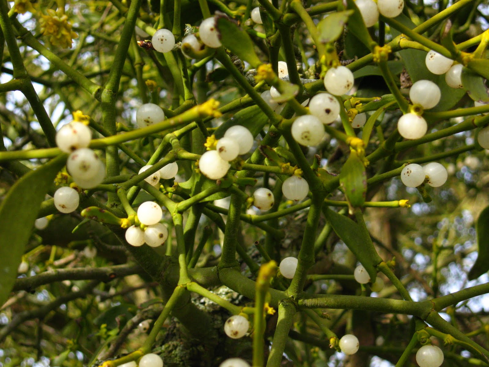 A cozy fire burning in the hearth, carols tinkling in the background, a glass of spiked eggnog in your bellies...what could be more romantic than finding yourself underneath a sprig of mistletoe hung in the archway with a sweetie, right? That is, if you don't mind kissing under a parasite. Viscum album, the European mistletoe is a parasite of more than 200 different trees and shrubs. It is hemiparasitic in that it still possesses chlorophyll but relies on its host plant for both water and other nutrients. Birds eat the juicy berries and then the seeds pass out in their feces and stick to the branch that they happen to land on, where they will germinate and set up a new parasitic plant. Mistletoe can severely damage or even kill their hosts if the infection is intense enough. The connection to Christmas is fairly recent, thought to have begun in the 18th century, and has evolved from a symbol that protected the home from fire to the more amorous excuse to kiss a pretty girl who happens to walk under it. This species, with its white berries, is more popular in Europe (and in plastic varieties everywhere); other mistletoe species (all also parasites) are used in other parts of the world.
December 16, 2010
December 16 - Desmozoon lepeophtherii
 Back in July, you met Lepeophtheirus salmonis, now meet Desmozoon lepeophtherii the hyperparasite that makes a living by infecting that particular parasitic copepod. Desmozoon lepeophtherii is a microsporidian, a diverse group of unicellular parasites that are the sister group to the fungi. Microsporidians infect a wide range of animal hosts, thus it is not surprising that even a parasitic copepod is not off-limits. Interestingly, genetic analyses indicate that the closest relatives of D. lepeophtherii are microsporidian in the genus Nucleospora, which are mostly parasites of salmonids. It is possible that for some reasons, the ancestor of D. lepeophtherii opportunistically made the jump from infecting its original fish host to infecting the ectoparasites which infects the said fish.
Reference:
Freeman, M. A. and Sommerville, C. 2009. Desmozoon lepeophtherii n. gen., n. sp., (Microsporidia: Enterocytozoonidae) infecting the salmon louse Lepeophtheirus salmonis (Copepoda: Caligidae). Parasite and Vector 2:58.
Contributed by Tommy Leung.
December 15, 2010
December 15 - "Blastocystis hominis"
 Single-celled organisms are difficult to classify. They don't have very much when it comes to morphology and so for a long time were just put into the large, amorphous group called "Protozoa" and treated as descending from a common ancestor. We now know that they are very divergent groups and today's parasite is a perfect example of the challenges of taxonomy. Blastocystis was originally thought to be closely related to yeasts, but then was moved to a large group called Sporozoa, which includes many well known parasites. DNA sequence data have shown, though that these parasites are part of another group known as the stramenopiles, which includes the diatoms, brown algae and Phytophthora infestans, the cause of Irish potato blight. Species of Blastocystis were classified based on the host that they had been found in, hence Blastocystis hominis. However, genetic studies showed that there is not a single species that infects humans, but rather nine or ten different subtypes, which have not yet been formally described (thus the quotation marks on the name.) Even more confusing than the taxonomy is the pathology. The protists live in the GI tract and are thought to produce typical types of GI-tract symptoms (do I have to list them?), but the symptoms reported are extremely varied and not everyone that tests positive for it shows symptoms.
December 14, 2010
December 14 - Fasciola hepatica
 This parasite can be baaaaad, to sheep - and to humans. Fasciola hepatica, or the common liver fluke, is a trematode parasite with a typical complex life cycle like so many that we have seen here before, involving snails that are common around pastures. Metacercariae are ingested by grazing animals and then they seek out liver tissue and feed for a month or two, causing anemia and other symptoms in the animal. Eventually, they mature into adults and settle down in the bile ducts and just churn out eggs - about half a million a day! Humans can become infected with this parasite through accidental ingestion of the metacercariae in water, on water plants, through contact with livestock and perhaps from ingesting raw liver from infected animals.
December 13, 2010
December 13 - Eudynamys scolopaceus
 This beautiful bird, commonly known as the Asian Koel, is a brood parasite in the cuckoo family. Eudynamys scolopaceus is found in southern Asia and primarily uses crows as hosts for its young. Although the female will sometimes remove one of the host's eggs when she lays hers, unlike many other brood parasites, the Asian Koel young usually do not kill or evict their nest mates, but rather beg their host parents in calls resembling those of baby crows. Their common name comes from the calls that the adults make, though their Sanskrit name, seen in literature that is more than 4000 years old, is Anya-Vapa which translates to "that was raised by others". Thus, the Asian Koel may be the first documented brood parasite known.
December 12, 2010
December 12 - Balamuthia mandrillaris
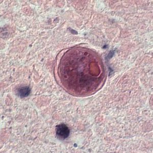 Normally, Balamuthia mandrillaris is a free-living amoeba, but on rare occasions, it has opportunistically become a parasite, with almost always fatal results. These single-celled organisms are thought to enter the body through either open wounds or perhaps inhalation and then they make their way to the brain, where they cause a disease condition called granulomatous amoebic encephalitis. Luckily cases of B. mandrillaris are extremely rare, but on the other hand, because they are rare, not much is known about their biology.
Image is from the CDC image library.
December 11, 2010
December 11 - Pelecitus fulicaeatrae
 Filarial worms are nematodes that typically inhabit the body cavities and subcutaneous spaces of vertebrates. Most are viviparous (live-bearing) and their larvae, called microfilariae are found in the blood stream or subcutaneous space, where they are drawn up by biting insects when they feed. When the insects feed on a second host, the microfilariae are transferred and the life cycle starts again. Pelecitus fulicaeatrae is found in the leg joints of waster birds (coots and grebes) and its intermediate host is a louse, one more case of a parasite being the intermediate host of another parasite.
Contributed by Mike Kinsella and photo by Julia Diaz.
December 10, 2010
December 10 - Parvatrema margaritense
 Trematodes are famous for the complexity of their life cycles, but some species demonstrate something beyond all expectations. A good example is Parvatrema margaritense which belongs to Gymnophallidae, a relatively small family of trematodes circulating in coastal ecosystems. As studied in the Bartents and White Seas in Russia, sexual reproduction of this parasite occurs in the digestive tract of the common eider duck (Somateria mollissima). Eggs containing miracidia disperse into the water and the bivalves Turtonia minuta get infected. The cercaria develop in sporocysts, are shed and then seek out and penetrate into the second intermediate host, the gastropod Margarites helicinus. The cercariae occupy the extrapallial cavity of their hosts, lose their tails and form metacercariae. For the majority of trematodes, the latter would eventually be infective for the definitive hosts. However, P. margaritense’s metacercariae are parthenogenetic; they produce the next generation of metacercariae, also parthenogenetic. These subsequently yield mature metacercariae that can continue the cycle inside the eiders. (In addition to these findings, other gymnophallid metacercariae from Sakhalin and the Kuril Islands in Russia were shown to produce cercariae which are capable of re-infecting the host species they originate from, so creating a peculiar sub-cycle).
One cercaria established in M. helicinus can thus give rise to as many as 1.5 – 2 thousand invasive metacercariae (clones). This is about 100 times as many as the number of cercariae shed by a single T. minuta a day, providing a significant multiplication of a parasite. Another curious observation is that metacercariae in the extrapallial cavity of M. helicinus don’t cause any of the common parasite-related troubles and might better be call commensals. The broader exciting discussion on these topics as well as on their evolutionary implications can be found in the referred papers.
References
Galaktionov K. V. An experimental study of the unusual life cycle of Parvatrema sp. (Trematoda: Gymnophallidae). Parazitologiya (1996), 30, 487–494 (in Russian).
Galaktionov K.V. Phenomenon of parthenogenetic metacercariae in gymnophallids and aspects of trematode evolution. Proc. Zool. Inst. Russ. Acad. Sci, 310. 2006: 51-58.
Galaktionov K.V., Irwin S.W.B., Saville D.H. One of the most complex life-cycles among trematodes: a description of Parvatrema margaritense (Ching, 1982) n. comb. (Gymnophallidae) possessing parthenogenetic metacercariae. Parasitology (2006), 132: 733-746.
Galaktionov K. V. A description of the parthenogenetic metacercaria and cercaria of Cercaria falsicingulae I larva nov. (Digenea: Gymnophallidae) from the snails Falsicingula spp. (Gastropoda), with speculation on an unusual life-cycle. Systematic Parasitology (2007), 68 (2), 137-146.
Contributed by Anya Gonchar.
December 9, 2010
December 9 - Cancellaria cooperi
 On this blog, we've had all kinds of blood-suckers - leeches, bats, ticks, and lice. But when it comes to vampirism, a snail doesn't usually come to mind, but that's exactly what today's parasite is - a blood-sucking snail. Cancellaria cooperi is a snail that appears to have specialised to feed on the blood of the Californian Torpedo Ray, Torpedo californica. These snails spend most of their time inactive, buried in the sand and waiting for the next potential victim. But when a torpedo ray comes along, this mollusc springs into action. Equipped with an extremely keen sense of smell, C. cooperi is capable of detecting the slightest trace of ray mucus, and observations of trails left by these blood-thirsty snails indicate that they can sniff out a ray from as much as 24 metres (about 80 feet) away. Upon making contact, the snail begin touching and exploring the dorsal surface of the ray with extended tentacles, before extending its proboscis and making a small incision with its scalpel-like radular teeth. It then insert its proboscis into the wound and begin its blood-sucking session, which can last for up to 40 minutes. This snail appears to be a specialised parasite of the California torpedo ray, and has no interest in approaching other benthic fishes which are common in its local area, though they have been observed to feed on the Angel Shark (Squantina californica) in laboratory settings. Surprisingly, the torpedo ray seems unperturbed by the experience of being felt up by a snail before getting cut and probed and having its blood-sucked by the vampiric mollusc. But then, torpedo rays seems to be generally unresponsive to most forms of prodding and mechanical stimuli.
 Source: O'Sullivan, J.B., McConnaughey, R.R. and Huber, M.E. (1987) A blood-sucking snail: the Cooper's Nutmeg, Cancellaria cooperi Gabb, parasitizes the California Electric Ray, Torpedo californica Ayre. Biological Bulletin 172: 362-366.
Post by Tommy Leung and photo by Lovell & Libby Langstroth.
December 8, 2010
December 8 - Sarcotaces arcticus
 Scarcely resembling a crustacean so much as some bloated, otherworldly maggot, Sarcotaces arcticus is virtually invisible when it swims up the backdoor of its preferred host, the rockfish. Attaching like a tick to the delicate membranes of the rectum, it begins to gorge on blood and releases an enzyme that causes the host's own body to grow a protective bag or "gall" around the intruder. A bag made of anus. Now impossible to dislodge, the parasite freely grows to around the size of a golf ball, which would be bad enough if the host were our size, but we're talking about a somewhat smaller fish here. Not only does the parasite make a sleeping bag out of rectum skin, it grows too large to ever fit back out either way. All the while, the parasite reproduces and releases its microscopic young, making it not only rude for the host to break wind at social gatherings, but positively terrifying.
Post by Jonathan Wojcik (of BogLeech fame - check out his cool parasite gear) and photo by Jonathan Martin (and if you're brave, check out the video that he took as well!).
December 7, 2010
December 7 - Trypanosoma cruzi
 Years after returning from the long voyage of the Beagle, Charles Darwin started to feel not-so-well - dizziness, muscle spasms, fatigue and other symptoms. Some speculate that the naturalist may have acquired Chagas Disease while he was traveling in South America. This malady is caused by a single-celled parasite known as Trypanosoma cruzi. These parasites are transmitted by triatomine bugs such as Rhodnius prolixus, commonly called "assassin bugs" or "kissing bugs", the latter because of their tendency to take blood meals around the mouth. The parasites are passed out in the bug's feces and enter the bite wound where they travel through the blood and can take up residence in numerous organs. The acute form may go unnoticed and the chronic form can take decades to set in. Infected people often die of heart failure, due to the damage done to this organ. Animals such as opossums, bats, and livestock serve as reservoirs for the parasites. In 2009, a paper documented transfer of genes from the trypanosome to humans followed by inheritance in the children of these Chagas Disease patients!
December 6, 2010
December 6 - Scaphanocephalus expansus
 One way to look at parasites is as generalists or specialists. Generalists are non-host specific and while some infect a group of related hosts, others may infect completely unrelated hosts. Specialists infect only one species of host. This strange-looking trematode with wing-like expansions on the anterior end is an intestinal parasite of the osprey, Pandion haliaetus. The osprey is so highly evolved that it is the only species in its own family, Pandionidae. Perhaps because of its evolutionary isolation, the osprey has an unusual number of helminth specialists, and because its diet is 99% fish, almost all of them probably use fish as an intermediate host.
Contributed by Mike Kinsella.
December 5, 2010
December 5 - Haemophilus influenzae
 Did you get a flu shot this year? Good. But, it's not going to protect you against this pathogen, Haemophilus influenzae. This gamma proteobacterium, a member of the Pasteurellaceae, was mistakenly thought to be the agent responsible for influenza until the 1930's when the actual culprit, viruses, were found. That said, these bacteria can still cause a whole slew of illnesses such as lower respiratory tract infections, pneumonia, ear infections, and meningitis. H. influenzae also holds another important distinction - it was the first bacterium to have its entire genome sequenced - this was published in 1995.
December 4, 2010
December 4 - Megarhyssa macrurus
 Megarhyssa macrurus is a species of parasitoid wasp. These insects seek out their hosts - larval horntail wasps - by tapping trees with their antennae until they sense their vibrations and scent. The females then bore into the wood with enormous ovipositors (now thought to be tipped with metals such as zinc!) and then she injects an egg into the larvae. Her offspring will hatch out and consume the body of its host and then use similarly metal-laden mandibles to emerge from the wood. Male M. macrurus are wandering around trees listening as well - they seek out newly emerging females to mate with.
December 3, 2010
December 3 - Schistosoma mansoni
 Schistosoma mansoni is an unusual parasitic trematode of humans. Unusual, because unlike most other trematodes, schistosomes are dioecious, i.e. have separate sexes and also unusual in that they are longer and more worm-like than most other flukes. Eggs are released in the host's feces and hatch into miracidia, which go on to infect freshwater snails and reproduce to form cercaria. The cercaria then seek out human hosts and penetrate their skin and make their way to the blood stream. The worms seek out a member of the opposite sex and if one is found, the pair will settle down in mesenteric blood vessels and begin to start a family. The couple is monogamous - the female actually lives within a groove in the male's body called the gynaecophoric canal (as shown in photo) and there she will churn out more than 300 eggs a day. S. mansoni is a major public health threat in parts of Africa, Asia, and South America where is causes a chronic disease known as either schistosomiasis or bilharzia.
December 2, 2010
December 2 - Myleusnema bicornis
 Myleusnema bicornis is a species of parasitic nematode belonging to the family Kathlaniidae which is found in the intestine of a small herbivorous freshwater fish call Pacoucine (Myleus ternetzi). This nematode has a few morphology features which are rather unusual. Firstly, while most nematodes have a relatively straightforward-looking anterior end, M. bicornis has a separate, narrow cephalic region that can be extended or retracted (see photo), superficially resembling the proboscis of acanthocephalans such as Profilicollis altmani and Echinorhynchus salmonis. Additionally, male M. bicornis worms have a pair of postcloacal horns located at the posterior end, a feature that is absent in all other species of nematode in the kathlaniid family.
Source: Moravec, F. and Thatcher, V.E. 1996. Myleusnema bicornis gen. et sp. n. (Nematoda: Kathlaniidae), an intestinal parasite of a freshwater serrasalmid fish, Myleus ternetzi, from French Guiana. Folia Parasitologica 43:53-59.
Contributed by Tommy Leung.
December 1, 2010
December 1 - Crossobothrium antonioi
 Crossobothrium antonioi is a species of tapeworm that was just described last year after finding it in the broadnose sevengill shark (Notorynchus cepedianus), off the coast of Argentina. This tapeworm may win the worldwide contest for having the most "cojones" as the colloquial saying goes - A mature worm can have over 60 segments or proglottids, and in every single one of them, C. antonioi has over 700 testes! That sounds rather formidable - until you realize that the entire tapeworm is only about 50 mm long.
November 30, 2010
November 30 - Pneunonema tiliquae
 Our parasite for today is a nematode called Pneunonema tiliquae and it is the only species within its genus. It is found in the lungs of the Eastern blue-tongue lizard (Tiliqua scincoides), a cute-looking skink from Australia which can grow to 30 cm (about a foot) long or more. Nothing is known about this parasite's life-cycle or how it enters the host. Based on what is known about other species of lung-dwelling nematodes in reptiles, it is likely that the blue-tongue lizard becomes infected through the oral route, when infective larvae in the environment are accidentally ingested by the lizard alongside its food.
A second parasite found by Tommy Leung in a roadkill skink he found. Click here to see the first.
November 29, 2010
November 29 - Equinurbia blakei
 The scanning electron microscope has allowed us to see the awesome symmetry of some nematodes up close and personal. Equinurbia blakei is an intestinal parasite of African elephants and one of a group called “large strongyles.” This group is characterized by complicated mouthparts called the corona radiata (radial crown) seen here. The four structures outside the crown are called amphids and are sensory organs. Large strongyles are characteristic of ruminants and some hosts, such as zebras, can harbor up to 20 species and over a million total worms per animal.
Contributed by Mike Kinsella.
November 28, 2010
November 28 - Halipegus eccentricus
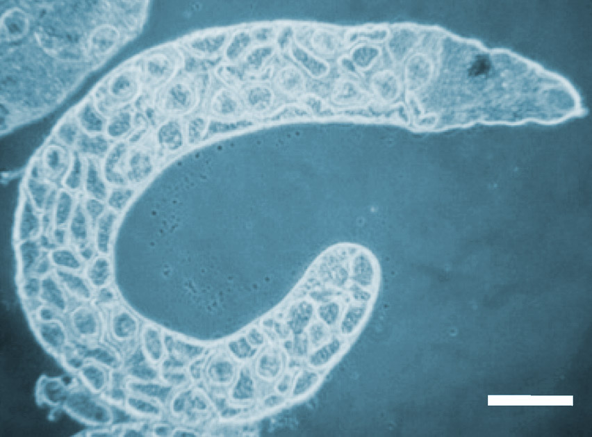 Halipegus eccentricus is a trematode parasite of North American frogs such as the bullfrog, Rana catesbeiana. Like many of the other trematodes we have met, H. eccentricus has a complex life cycle involving many different hosts. The first intermediate host is a snail of the genera Physa or Planorbella. The cercariae are then ingested by the second intermediate host, a small crustacean, such as a copepod or an ostracod. The metacercariae were then thought to be ingested by tadpoles where they waited for the amphibian to develop into a mature frog, at which point, the parasite would migrate to the frog's eustachian tubes (yes, these worms live in frog ears.) A recent study, however, showed that odonate insects (damselflies, dragonflies) serve as paratenic hosts for the trematodes and that only adult frogs are becoming infected. We are always learning more about parasites!
The image comes from the paper above and shows a redia of H. eccentricus, with the minute cercaria developing inside.
November 27, 2010
November 27 - Fregata minor
 Imagine someone waiting until just after you've swallowed your last bite of Aunt Tillie's famous pumpkin pie this Thanksgiving, and then forcing you to run a marathon until the inevitable gastric upheaval. Then, without as much as a "thank you", they catch the product of your upheaval, eat it, and run away. Shamelessly disgusting! This is how the Great Frigatebird (Fregata minor) makes a portion of its living, as an "on-the-wing" kleptoparasite. This Blog has featured "kleptos" before, but none as disgustingly brash and acrobatic as the Frigatebird. This enormous (2 meter wingspan) iridescent black seabird literally harasses (often at breakneck speeds) other self-respecting piscivorous birds until they "toss their fish-sticks". It doesn't stop here; because this bird cannot take off without a freefall (an evolutionary compromise of having the highest wing-size to body-size ratio of any bird) it cannot land on the water to eat its meal. Instead it has to perform amazing acrobatics to "catch the retch" before splashdown. When not "downing up-chuck" it feeds on flying fish caught "on-the-wing". Frigatebirds are also interesting in many less vile ways, not the least of which is the fact that they look a little like pterodactyls!
Post and photo by Matt Leslie.
November 26, 2010
November 26 - Genarchella astyanactis
 Today's parasite - Genarchella astyanactis - belongs to family of digenean trematodes called the Derogenidae, which have evolved a unique way of infecting their second intermediate hosts. They use copepods and other small crustaceans as second intermediate hosts, and their strangely-shaped cercariae are propelled from the snail first intermediate host by a forked appendage. The cercariae are also armed with a coiled structure call a "delivery tube" (shown extended in the drawing) that they use to gain access to the copepod host. For some reason, copepods identify the strange cercaria as a food item and avidly seize any that they encounter. However, as the crustacean manipulates the parasite with its mouth parts, the delivery tube of the cercaria rapidly extends and penetrates the copepod's gut wall. At the same time the cercaria body is propelled through the delivery tube into the body cavity. Imagine eating a muffin which stabs the back of your throat the moment that you bite it, and then injects a squirming parasite into your body!
Reference:
Ditrich, O., Scholz, T., Aguirre-Macedo, L. and Vargas-Vázquez, J. 1997. Larval stages of trematodes from freshwater mollusc of the Yucatan Peninsula, Mexico. Folia Parasitologica 44:109-127.
Contributed by Tommy Leung.
November 25, 2010
November 25 - Chelopistes meleagridis
 Happy Thanksgiving to all the U.S. readers out there! Seemed appropriate to feature another turkey parasite today (we've had two others recently, i.e. Syngamus trachaea and Trichomonas gallinae ), but this one is something that anyone whose exposure to turkeys is limited to defrosting a Butterball will have no opportunity to come into contact with: the Large Turkey Louse, Chelopistes meleagridis. These lice are very common, especially on wild turkeys and have been introduced to many places via transport of their avian hosts.
November 24, 2010
November 24 - Lemurpediculus verruculosus
 Lemurpediculus verruculosus is a species of louse that infects Microcebus rufus, commonly known as the Eastern Rufous Lemur or the Brown Mouse Lemur. L. verruculosus has a penchant for areas of its host where the skin is thin, the peripheral blood supply is good and where the animal cannot easily groom - thus the ears and, if it is on a male, the testes. This species was originally discovered back in 1951 as part of a collecting expedition by the famous medical entomologist Harry Hoogstraal, but the species description was based only on a single female that had been collected. Recent work on mouse lemurs yielded many more specimens of these lice, allowing the description to be expanded to include males and instar stages as well.
 Photo contributed by Lance Durden, one of the authors of the new paper.
November 23, 2010
November 23 - Transvena annulospinosa
Reference:
Pichelin, S. and Cribb, T.H. (2001) The status of the Diplosentidae (Acanthocephala: Palaeacanthocephala) and a new family of acanthocephalan from Australian wrasses (Pisces: Labridae). Folia Parasitologica 48: 289-303.
Contributed by Tommy Leung.
November 22, 2010
November 22 - Trichomonas gallinae
 There's certainly no doubt that dinosaurs had parasites. The problem is, soft-bodied things like tapeworms and nematodes, let alone smaller things like trypanosomes or malaria parasites just don't fossilize well, so actually being able to say which species of parasites the "terrible lizards" might have been infected with is close to impossible. Recently, though, jaw bones from Tyrannosaurus rex specimens were re-examined and lesions in them were attributed to a parasite that continues to plague modern-day birds, Trichomonas gallinae. These single-celled parasites, closely related to the human STD, Trichomonas vaginalis, produce "cheesy" lesions in the mouth, pharynx and crop of birds such as pigeons, chickens, and your soon-to-be Thanksgiving turkey, causing a disease that is sometimes called "canker". These birds acquire the infection through consumption of contaminated water, but avian predators, such as falcons, can also be infected from eating parasitized prey. The authors of the paper that makes this link between lesions and T. gallinae hypothesize that even the Field Museum's famous "Sue" may have died of starvation as a result of this parasite damaging its mouth so badly. The image is an artist's vision of what the parasitized dino may have looked like. (Click on the thumbnail to get a better look and see the "cheesy" lesions in its mouth and check out this site for pictures of the parasite in pigeons.)
November 21, 2010
November 21 - Tetracladium sternae
 Although most digenetic trematodes (except for schistosomes) are hermaphroditic and thus, by definition, capable of self-fertilization, there is some evidence that cross-fertilization may be quite common and maybe even the norm. But it is unusual to catch trematodes in the act of fertilization. There is one exception - Tetracladium sternae is a trematode found in shorebirds such as terns and shearwaters and it is often found in copulating pairs.
Post and image by Mike Kinsella.
November 20, 2010
November 20 - Pedicularis groenlandica

Pedicularis groenlandica, or as it is commonly known as because of the shape of its flowers (see insert), Elephant's Head or Elephanthead Lousewort (a big handle for a little flower!) is a parasitic plant in the broomrape family (see also Boschniakia hookeri and Orobranche californica). These plants can be found in - you guessed it - Greenland, but also across Canada and into western North America. P. groenlandica uses haustoria to penetrate the roots of other plants and then suck their water and nutrients out.
November 19, 2010
November 19 - Asymphylodora tincae
 Today's parasite is commonly found in freshwater fishes of the Palaearctic region. This particular worm has evolved to skip a few steps to the usual three-host life-cycle of most digeneans. Asymphylodora tincae uses a snails as a first intermediate host, where larval stages known as cercariae are produced through asexual multiplication - so far so usual for digeneans. However, instead of leaving the snail like most digeneans, the tailless cercariae stay inside the snail. Additionally, instead of then developing into the cyst-like metacercariae stage as a prelude to infecting the definitive host, this species has done away with that altogether. The cercariae of A. tincae can directly infect the fish definitive host, which occurs when the fish consumes an parasitised snail.
Reference and photo source:
Našincová, V. and Scholz, T. (1994) The life cycle of Asymphylodora tincae (Modeer 1790) (Trematoda: Monorchiidae): a unique development in monorchiid trematodes. Parasitology Research 80:192-197.
Contributed by Tommy Leung.
November 18, 2010
November 18 - Clinostomum sp.
 This is a metacercarium of a Clinostomum species that was found encysted in the fin of Perca flavescens, the yellow perch. The definitive hosts of these trematodes are fish-eating birds and reptiles, and adult clinostomes are commonly found in the mouth and esophagus. Eggs of Clinostomum are shed in the feces, hopefully in the water. Miricidia then infect planorbid snails. Cercariae released from the snails penetrate the skin fish and amphibians (the second intermediate hosts), encysting as metacercariae throughout the body. Definitive hosts become infected when feeding on infected fish. Clinstomum metacercaria are often large and yellow in appearance, thus their presence is often called “yellow grub disease”.
 Contributed by Jessica Light.
November 17, 2010
November 17 - Tetrabothrius sp.
 The parasite for today is a tapeworm recovered from the intestine of an Andrews' Beak Whale (Mesoplodon bowdoini). This worm belongs to the genus Tetrabothrius, and while this particular species infects beaked whales (obviously), other species of Tetrabothrius have been found in a range of dissimilar marine hosts ranging from baleen whales to fish-eating birds such as albatross and penguins. Tapeworms like Tetrabothrius often have species-specific morphology of their scolex (which is the organ they use to attach themselves to the intestinal wall), and such features can be used to distinguish different species. Larval stages of tapeworm often lack such distinguishing characteristics, making their identification practically impossible. However, the advent of molecular biology technique has enable scientists to use DNA sequences from the larval stages and match them up to those taken from adult worms, allowing their full life-cycle to be mapped out.
Contributed by Tommy Leung.
November 16, 2010
November 16 - Opisthorchis viverrini
 Stay out of the sun to avoid skin cancer. Don't smoke to avoid lung cancer. Don't eat raw fish to avoid liver cancer? It sounds a little strange, but in fact, the liver flukes Clonorchis sinensis and Opisthorchis viverrini have been found to be carcinogenic, inducing a cancer of the bile ducts that is very often fatal. The life cycle of O. viverrini is very similar to C. sinensis - they first pass through snails, then fish, and then finally the adults inhabit mammals that like to eat fish, including cats, dogs, and humans. The region of Khon Kaen, in northeast Thailand, has the highest incidence of these bile-duct cancers in the world - and 70% of the people in this region are infected with O. viverrini. A recent study produced large libraries of sequences of the transcriptomes - the genes that are transcribed at various stages - of these two important parasites, thus it is hopeful that new drug targets can be identified.
 You can read more about the fluke/cancer link here.
November 15, 2010
November 15 - Pharyngodon australis
 I was on my way home from grocery shopping when I spotted something in the middle of the road near where I live. As I got closer I saw that it was a dead lizard. So like any good parasitologist, I quickly got home, parked my car, grabbed some plastic bags and dashed across the road, scooping up the lizard in the process. It must have only just been recently killed because rigor mortis hasn't even set in. So I thought I'd make something worthwhile out of an otherwise senseless death, drove to work and started dissecting the dead lizard, and sure enough, found this parasite! Pharyngodon australis is a species of nematode found in the large intestine of Eastern blue-tongue lizard (Tiliqua scincoides), a large ominvorous skink from Australia. Thousands of nematodes live in the gastrointestinal tract of skinks and other lizards. Stable isotope studies have indicate that some of these nematodes might be consuming microbes living in the host's gut, while other experiments showed that they might even contribute to gut fermentation. So this might be a case of what would normally be assumed to be a parasitic organism actually being a welcome guest!
Contributed by Tommy Leung.
November 14, 2010
November 14 - Pseudolynchia canariensis
 Pseudolynchia canariensis is a hippoboscid or louse fly that feeds on pigeons and doves and can transmit the blood parasite Haemoproteus columbae. This species is primarily found in Africa and Asia. If you click on this photo of a P. canariensis fly and look at it carefully, you can see tiny little pink dots near the back of its abdomen. These even smaller things are mites, though in this case they are not really parasitic, but rather phoretic, a phenomenon whereby one kind of organism uses another as a means of transportation. All aboard! This fly is now departing...
November 13, 2010
November 13 - Brugia malayi
 Like Wuchereria bancrofti, Brugia malayi is another type of filarial nematode that is responsible for the disease known as lymphatic filariasis or, as it is better known, elephantiasis. The name comes from the fact that the adults inhabit the lymphatic system and their presence induces intense swelling, such that the skin resembles that of a saggy elephants. This species is found in Southeast Asia, including Vietnam, China, India, and other neighboring countries. It's estimated to infect over 13 million people in this part of the world. Prevention and treatment strategies include vector control and drugs, some of which are being newly developed thanks to the availability of a complete genome sequence.
November 12, 2010
November 12 - Amphibiocystidium ranae
 Today we are featuring a fungal parasite of frogs. This parasite is former known as Dermocystidium ranae and was classified within a genus which also included a number of fungal parasites of fishes. However, further research on D. ranae found that it has a number of life-cycle and morphological features which separate this parasite from others within the Dermocystidium genus. Because of those distinguishing characteristics, it has now been reclassified and placed in a genus of its own - Amphibiocystidium - to reflect its unique status. While the parasite in the fish-infecting sister genus, Dermocystidium, has recieved much scientific interest over the last 50 or so years, far less research has been conducted on Amphibiocystidium ranae and it is not clear if it cause any actual harm to its amphibian host or if it is more of a benign parasite.
Reference:
Pascolini, R., Daszak, P., Cunningham, A.A., Tei, S., Vagnetti, D., Bucci, S., Fagotti, A. and Di Rosa, I. (2003) Parasitism by Dermocystidium ranae in a population of Rana esculenta complex in Central Italy and descriptiion of Amphibiocystidium n. gen. Diseases of Aquatic Organisms 56: 65-74
Contributed by Tommy Leung.
Subscribe to:
Posts (Atom)